
See the updates at the bottom. As of November, 2017, we now have Allen Steere, Gary Wormser and Linden Hu all admitting in separate reports that the fungal antigen Osps (or Vmps, and probably glycolipids) that are shed from and make up Borreliae are responsible for the severity of this IMMUNOSUPPRESSION disease, as TLR2/1 agonists.
(Note added Nov 8, 2017)
KEY CONCEPT: “Modulation of the apoptotic machinery during viral and bacterial infections, as well as in various malignancies, is a well established mechanism that promotes the survival of affected cells.”
Both lipoproteins of the OspA type shed by these Lyme “stealth bombers” and Epstein-Barr inhibit apoptosis. And they both hang out in lymph nodes and B cells. Think about it.
Other Key Concept: There is more DNA (plasmid) packaged in released blebs (or vesicles or exosomes) – which have fungal osps all over them – than there are spirochetes. Spirochetes are “stealth bombers.” That is how they cause chronic or immunosuppression disease.
DNA Is Packaged within Membrane-Derived Vesicles of GramNegative but Not Gram-Positive Bacteria
“Recently, DNA packaged within nuclease-resistant membrane vesicles of Neisseria gonorrhoeae andBorrelia burgdorferi was described. This study assayed 18 species of gram-negative and gram-positive eubacteria for nuclease-protected DNA associated with extracellular membrane vesicles. Vesicles from only the gram-negative bacteria contained nuclease-protected linear or supercoiled DNAs or both.”

http://www.ncbi.nlm.nih.gov/pmc/articles/PMC184538/pdf/aem00087-0464.pdf
=====================================
There is more DNA (plasmid) packaged in released blebs – which have fungal osps all over them – than there are spirochetes. Spirochetes are “stealth bombers.” That is how they cause chronic or immunosuppression disease:
J Infect Dis. 1994 Mar;169(3):668-72.
Target imbalance: disparity of Borrelia burgdorferi genetic material in synovial fluid from Lyme arthritis patients.
Persing DH1, Rutledge BJ, Rys PN, Podzorski DS, Mitchell PD, Reed KD, Liu B, Fikrig E, Malawista SE.
“Lyme arthritis is a late manifestation of Lyme disease that results in episodic synovial inflammation and swelling. Although this process is thought to be driven directly by the spirochetal etiologic agent, Borrelia burgdorferi, the organism itself has been recovered by culture only twice. In contrast, polymerase chain reaction (PCR) studies are usually positive. This apparent discrepancy in 19 culture-negative synovial fluid specimens from 18 patients with Lyme arthritis was investigated. In all 19, DNA sequences characteristic of plasmid-encoded genes OspA and OspB were easily detected. However, despite equivalent or even superior analytic sensitivity for detection of cultured organisms, the reactivity of two genomic DNA targets was often weak or absent altogether in the clinical specimens. This apparent overrepresentation of B. burgdorferi plasmid sequences was found exclusively in clinical specimens and not in cultured organisms. The physiologic imbalance of genomic and plasmid DNA reactivity in B. burgdorferi infection may signal an underlying pathogenetic mechanism.http://www.ncbi.nlm.nih.gov/pubmed/8158048“
Alan Barbour on “Stealth Bombers”
“….Though most cases of B. burgdorferi infection can be eliminated easily by a short course of antibiotics, Lyme disease can be disabling if left untreated. Scientists are searching for answers to how the relatively small numbers of bacteria (1,000 microbes per milliliter in infected humans) can cause excessive inflammation, and why the spirochetes localize to certain tissues, including the joints, the heart, and the brain. They suspect the key to the mystery lies on the spirochetes’ surface, in the collection of proteins that coat its outer membrane.
“Patients with Lyme disease experience flu-like symptoms-aches, fever, nausea, joint pain. When the spirochetes first get under the skin, they mark their point of entry with a characteristic bull’s-eye rash. Left unchecked, the B. burgdorferi bacteria will work their way into the bloodstream and head off to other organs. In late-stage Lyme disease, a month or two after infection, the spirochetes take up residence in the heart or the joints, where they cause inflammation. Others settle in the brain, where the body’s immune system can’t reach them as easily.
“Working with the related spirochete B. turicata, Barbour has found that bacteria that wind up in the brains of immune-deficient mice differ by only a single protein from the bacteria that remain in the blood. The protein, which shows homology to the outer-surface protein OspC from a variety of B. burgdorferistrains, may allow the spirochete to establish the kind of long-lasting brain infection that underlies lingering Lyme disease, he theorizes.
“According to Barbour, B. burgdorferi lacking its outer-surface proteins (including OspA, OspB, OspC, and OspD) is easily killed by the blood’s complement system. Several groups of researchers are currently characterizing which proteins B. burgdorferi loses when it is kept in culture conditions for too long. When the spirochete is raised without contact with a mammalian host, it is unable to infect mice. And, according to microbiologists James Carroll and Frank Gherardini of the University of Georgia, it may also lose OspD and a handful of previously unidentified surface proteins (J.A. Carroll, F.C. Gherardini, Infection and Immunity, 64:392-8, 1996).
“Many researchers believe that the secret to B. burgdorferi‘s infectivity and inflammatory capacity lies in the interaction of its surface proteins with the host’s immunological system. Yale researcher Stephen Barthold, a veterinarian and professor of comparative medicine who developed the first mouse model of Lyme disease, studies the expression of B. burgdorferi surface proteins throughout various stages of the spirochete’s life cycle. He finds that during the early stages of infection, B. burgdorferiavoids immune detection by decreasing its expression of surface proteins or cloaking its expressed surface proteins under a layer of slime. “It’s using some sort of stealth-bomber-type mechanism,” he says. Or, using another diversionary tactic called blebbing, the spirochete can pinch off bits of its membrane in order to release its surface proteins. Explains Barbour: “It’s like a bacterial Star Wars defense program,” in which released surface proteins might intercept incoming host antibodies, keeping the spirochete safe from immunological attack.
 TINY CREATURES: In size order, the three tick feeding stages are nymph, larva, and adult, shown next to a U.S. dime. |
During the early stages of infection, perhaps while the spirochete is still inside the tick, B. burgdorferi switches on a whole new family of surface proteins that may help it adapt to life in its mammalian host, according to Barthold. These so-called in vivo expressed antigens may help the spirochete invade tissues or elicit inflammation in the joints (S.W. Barthold et al.,Laboratory Investigation,74:57-67, 1996).
“Although they can offer educated guesses, researchers are still unsure what roles the surface proteins of B. burgdorferi play in infection and disease. A group of protein chemists and crystallographers at Brookhaven National Laboratory in Upton, N.Y., and the State University of New York, Stony Brook, are taking a more structural approach. John Dunn and Cathy Lawson purified and crystallized OspA-a surface protein that B. burgdorferi expresses when it lives in the midgut of the tick. The structure might offer some clues to how the protein functions, says crystallographer Lawson. Further, because the Lyme disease vaccines now being tested in humans contain OspA, Lawson says, “it would be nice to know what the protein looks like.” The researchers presented their results at the seventh International Conference on Lyme Borreliosis, held in San Francisco late last month. The biennial meeting draws more than 400 Lyme researchers from around the world….”
http://www.the-scientist.com/?articles.view/articleNo/17985/title/Researchers-Finding-Rewarding-Careers-As-Software-Entrepreneurs/
The shed blebs (or exosomes or vesicles) have LYMErix on them (delayed fuse or “time bomb”):
Characterization of multiprotein complexes of the Borrelia burgdorferi outer membrane vesicles.
“Although we uncovered the existence of at least 10 distinct OM complexes harboring several unique subunits, the complexome is dominated by the frequent occurrence of a limited diversity of membrane proteins, most notably P13, outer surface protein (Osp) A, -B, -C, and -D and Lp6.6.”
Many groups have tried to develop tests based on these shed antigens, including the NIH, and the NIH again recently, and the LUAT or Igenex’s Lyme urine antigen test. Most recently NIST and Johns Hopkins made this claim:
http://www.ncbi.nlm.nih.gov/pubmed/26491962
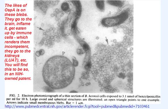
See for yourself, with your own eyeballs, what is the relative abundance of blebs vs spirochetes in regular culture condition (without antibiotics), and also with antibiotics. This is by Alan Barbour in 1982:
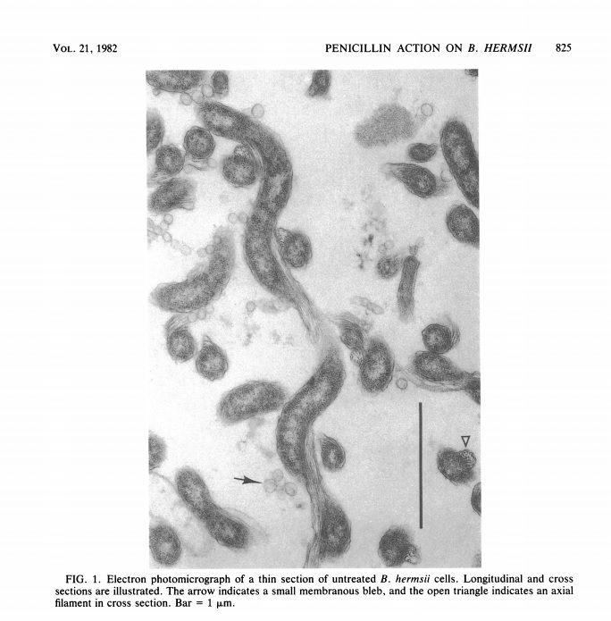
http://www.ncbi.nlm.nih.gov/pmc/articles/PMC182019/pdf/aac00017-0141.pdf
http://www.ncbi.nlm.nih.gov/pmc/articles/PMC182019/pdf/aac00017-0141.pdf
====================================
SEMANTICS GAME
Why do we say this is a semantics game? Because you think you are fighting with the CDC Lyme criminals over Persisting Spirochetes (of course they are permanent), vs. No Persisting Spirochetes. And even when spirochetal DNA is found in treated patients, the bad guys say, that is still not “Lyme disease.”
When someone has a legal case, say, for compensation because they were bitten on the job and became ill, these criminals will come and perjure themselves in court, and say “This person does not have Lyme disease,” (even if they had several bands and a witnessed tick bite rash).
They get away with committing perjury because “Lyme disease” means only one thing. ONLY ONE THING…. since the CDC pulled the Dearborn stunt in 1994….
“Lyme disease” is ONLY “a bad knee” and no other symptoms.
NO dementia,
no fatigue,
no neuritis or nerve pain,
no headache,
no double vision,
no sleep disorders,
no vasculitis,
no multiple-sclerosis, stroke, Lupus, ALS, or cancer outcomes.
“Lyme disease” IS NOT a “Great Imitator.”
It, “Lyme disease” is some research fraud that happened in Europe with Allen Steere and little Frankie Dressler – alone – at the same time CDC officers like Barbara Johnson were filing patents for recombinant vaccine and test kit products with SmithKline in Europe (1992-1993).
It used to be called Lyme relapsing fever or Lyme borreliosis, and historically, spirochetal diseases are incurable and devastating – not to mention chronic or a permanent disability – but “Lyme disease?” That’s something that happened in the minds of people who would not be able to earn royalties from this new DNA technology and the Bayh-Dole Act, where people can OWN DNA and therefore OWN diseases, UNLESS THEY LIED ABOUT IT, and acted like spirochetes were just regular bacteria and not their own ancient phylum, and shedder of triacyl lipoproteins or FUNGAL ENDOTOXINS!!
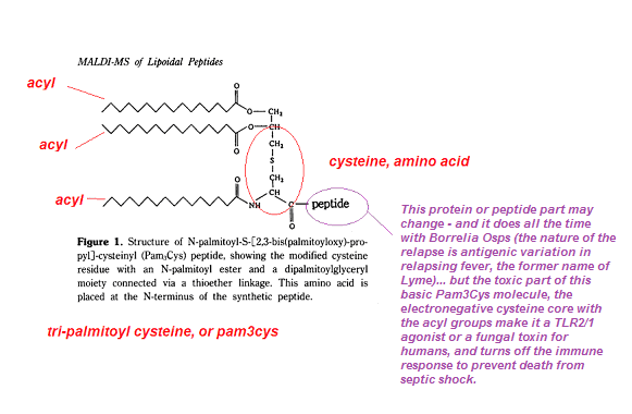
Pam3Cys is a triacylated lipoprotein, the degree of acylation is equated with its toxicity. So what is acylation? It’s the zig-zaggy lines that mean Carbon-Carbon-Carbon, yes, hydrocarbons, like margarine or octane. Exactly, the name just refers to the number of carbons in each carboxyl or acyl group. Palmitic (the Pam in Pam3Cys) has X number of carbons, gasoline, 8, linoleic acids, like 14. Look up what are alkanes then add a COOH group and you have one of these fatty acids.
Something highly acylated like this (3 or more fatty acids hanging off) are managed by Toll-like Receptor (TLR) 2 and TLR1, together. Therefore a “TLR2/1-agonist” is another term that generally refers to lipoproteins like those from Borrelia, mycoplasma, mycobacteria, and others like Brucella. (But they can manage other compounds.)
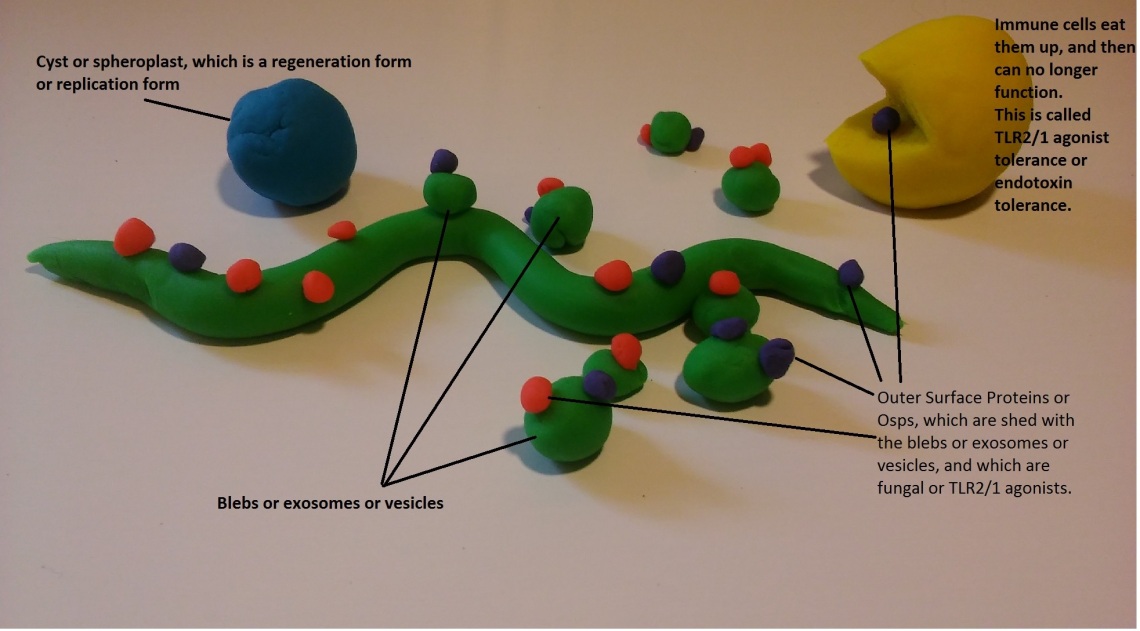
This thing, Pam3Cys and fungal lipid molecules like it, is shed with the blebs. In other words, like this:
What good would patented DNA be to people if everyone knew tick borne diseases, and especially borrelia spirochetes were an ancient phyla, not really “bacteria” in the normal sense, and most of all SHED FUNGAL ANTIGENS, like brucella and the mycos (tuberculosis, pneumonia, etc).
Go ahead and “ask your doctor” what myco means. ‘Good chance he has no clue, and never heard of immunosuppression diseases besides AIDS.
No, your “doctor” does not know about shedders of fungal antigens, does not know what anyone means when they say “fungal,” does not know any taxonomy, does not know how to use the internet, even, to look up what spirochetes are and do, and probably thinks rheumatology and even psychiatry are real things.

This ^^^ is “Lyme disease.” Bad knee. ONLY.
This is what happened at Dearborn. “Lyme disease” the definition, the “case definition serology” or the “2 tiered case definition” became “only a swollen knee and no other symptoms.”
Not says your’s truly, but Allen Steere, the CDC, and all the perjurers of the ALDF.com and IDSA who participated in the OspA vaccines trials and in falsifying the case definition in 1994 at Dearborn.
SO, WHAT IS THE OTHER THING, THEN, the thing left out of the Dearborn case definition and why do we keep harping on the herpes?
Paul Duray:
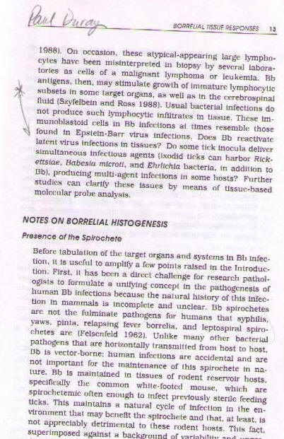
“On occasion, these atypical-appearing large lymphocytes have been misinterpreted in biopsy by several laboratories as cells of a malignant lymphoma or leukemia. Bb antigens, then, may stimulate growth of immature lymphocytic suibsets in some target organs, as well as in the cerebrospinal fluid (Szyfelbein and Ross 1988). Usual bacterial infections do not produce such lymphocytic infiltrates in tissue. These immunoblastoid cells in Bb infections at times resemble those found in Epstein-Barr virus infections. Does Bb reactivate latent virus infections in tissues? Do some tick inocula harbor simultaneous infectious agents (ixodid ticks can harbor Rickettsiae, Babesia microti, and Ehrlichia bacteria, in addition to Bb), producing multi-agent infections in some hosts? Further studies can clarify these issues by mans of tissue-based molecular probe analysis.” –
Paul Duray, NCI, NIH, Ft. Detrick, at the 1992 Cold Spring Harbor Crooks’ Conference, published in Steve Schutzer’s Lyme Disease: Molecular and Immunologic Approaches.
Now go look up what EBV-like transformed or immortalized cells means.
Duray:
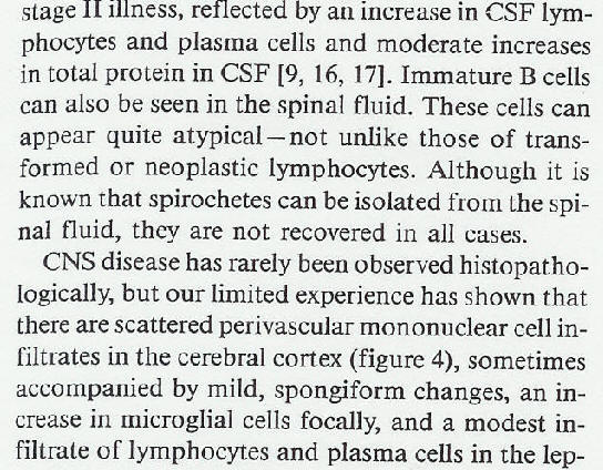
“Immature B cells can also be seen in the spinal fluid. These cells can appear quite atypical- not unlike those of transformed or neoplastic lymphocytes.” —
–1989, IDSA’s Journal
Now go look up what “EBV-like transformed” or “immortalized cells” means.
Now go look up what “EBV-like transformed” or “immortalized cells” means.
Do not pass go, do not collect 200 dollars. JUST go find out what I am talking about. And you better, because you know I’ll come after you on Facebook and call you stupid and lazy.
What else inhibits apoptosis of infected B cells?
2) BCL2 class genes (look that up too) and….
3) LIPOPROTEINS:
NF-kappaB was transactivated in cells treated with M. fermentans lipoproteins, and was essential for host cell survival, but not for the inhibition of TNFalpha-induced apoptosis by LPMf. Our results suggest that the inhibitory effect exerted by M. fermentans on TNFalpha-induced apoptosis in U937 cells is due to the membrane lipoproteins of these bacteria. http://www.ncbi.nlm.nih.gov/pubmed/16889623
2009; Anti-apoptotic genes in the survival of monocytic cells during infection.
Macrophages are cells of the immune system that protect organisms against invading pathogens by fulfilling critical roles in innate and adaptive immunity and inflammation. They originate from circulating monocytes and show a high degree of heterogeneity, which reflects the specialization of function given by different anatomical locations. Differentiation of monocytes towards a macrophage phenotype is also accompanied by an increase of resistance against various apoptotic stimuli, a required characteristic that allows macrophages to accomplish their function in a stressful environment.Apoptosis, a form of programmed cell death, is a tightly regulated process, needed to maintain homeostasis by balancing proliferation with cellular demise. Caspases, a family of cysteine proteases that are highly conserved in multicellular organisms, function as central regulators of apoptosis. FLIP (FLICE-inhibitory protein), anti-apoptotic members of the Bcl2 family and inhibitors of apoptosis (IAP) are the main three groups of anti-apoptotic genes that counteract caspase activation through both the extrinsic and intrinsic apoptotic pathways. Modulation of the apoptotic machinery during viral and bacterial infections, as well as in various malignancies, is a well established mechanism that promotes the survival of affected cells. The involvement of anti-apoptotic genes in the survival of monocytes/macrophages, either physiological or pathological, will be described in this review. How viral and bacterial infections that target cells of the monocytic lineage affect the expression of anti-apoptotic genes is important in understanding the pathological mechanisms that lead to manifested disease. The latest therapeutic approaches that target anti-apoptotic genes will also be discussed.
M.tb also exploits TLRs to induce anti-apoptotic genes that enhance cell survival and promote bacterial persistence [109]. Exploiting TLRs is not a mechanism unique to M.tb. As mentioned earlier, we have shown that TLR3, TLR4 and TLR9, when stimulated by their ligands PolyI:C, LPS and CpG DNA, respectively, protected monocytic cells from HIV-Vpr induced apoptosis by induction of NFκB and anti-apoptotic cIAP genes (unpublished data). Stimulation of TLR2, found in abundance at sites of M.tb infection, by components of M.tb cell wall, has been shown to protect human macrophages against apoptosis. THP1-derived macrophages when stimulated with 19kDa mycobacterial lipoprotein or mannosylated LAM were shown to induce resistance to apoptosis via activation of NFκB and subsequent induction of anti apoptotic cFLIP which inhibits death receptor-mediated apoptosis [25, 109].
http://www.ncbi.nlm.nih.gov/pubmed/20119528
http://www.ncbi.nlm.nih.gov/pmc/articles/PMC2729995/?tool=pubmed
Mycobacterium bovis Bacillus Calmette Guérin infection prevents apoptosis of resting human monocytes.
Apoptosis plays an essential role in the development and homeostasis of multicellular organisms. Some infectious agents interfere with this programmed cell death to their own benefit. Here, we show that infection of resting human monocytes with Mycobacterium bovis Bacillus Calmette Guérin (BCG) increases monocyte viability by preventing them from undergoing apoptosis. Heat-killed BCG also prevented apoptosis, indicating that replication of BCG is not required to prevent cell death. Analysis of BCG-infected monocytes revealed an up-regulation of the A1 mRNA, whereas the bcl-2 mRNA was not up-regulated. Interestingly, preinfection with BCG renders the cells resistant to interleukin (IL)-10-induced apoptosis which may be one of the mechanisms mycobacteria use to modulate immune responses. BCG infection was also accompanied by an impairment of the capacity of monocytes to secrete IL-10 and by an induction of the capacity to secrete tumor necrosis factor-alpha, two cytokines known to induce and prevent human monocyte apoptosis, respectively. Since it has been reported that apoptosis is involved in killing of intracellular mycobacteria, the prevention of apoptosis may represent a strategy for mycobacterial survival in the infected host.
http://www.ncbi.nlm.nih.gov/pubmed/9341792?dopt=Abstract
http://onlinelibrary.wiley.com/doi/10.1002/eji.1830270945/abstract
What does Gary Wormser say about OspA and “blocking the cell phase cycle progression” in immune cells?
Gary Wormser saying – while LYMErix was still on the market -, that how sick you become, depends on how much OspA you got stuck with, either by ticks/spirochetes or syringe. You cant make this up:
“The magnitude of modulation [immunosuppression – KMD] was directly dependent on the quantity of OspA. OspA interferes with the response of lymphocytes to proliferative stimuli including a blocking of cell cycle phase progression.”
“We have previously demonstrated that proteins of B. burgdorferi are capable of modulating human cellular immune responses [7]. Suppression of in vitro mitogen- or antigen-mediated proliferative responses of lymphocytes and reduced production of interleukin-2 (IL-2) from lymphocytes were demonstrated using protein extracts of B. burgdorferi. These early studies were confirmed by a report of de Souza et al. [8], who observed that B. burgdorferi infection in mice resulted in impaired T and B cell proliferation to mitogens and reduced IL-2 and IL-4 production. The nature of the B. burgdorferi proteins responsible for suppression of cellular immunity has not been defined. In this study we examined the modulating activity of a recombinant outer surface protein A (OspA) vaccine preparation on cellular immune responses.”
http://femsim.oxfordjournals.org/content/28/3/193.long
Now, where do Borrelia like to live and where do EBV like to live, … now that you know both cause the inhibition of infected immune cells like B cells?
Right. In the lymph nodes. Together 😀
So, an EBV (et al) latently infected B cell comes in contact with Borrelia lipoproteins like OspA shed or EXPORTED by Borrelia and whaddya got?
Right, EBV (etc) become unlatent. Viola! >>> Great Imitators like Lupus, MS, cancer and all the other well known outcomes of NOT “LYME DISEASE.”
Let’s see if anyone has shown whether or not EBV and Lyme together is a productive marriage:
Interaction of Borrelia burgdorferi sensu lato with Epstein-Barr virus in lymphoblastoid cells.
”Since the possibility of interruption of latent EBV infection has been suggested by the induction of the lytic virus cycle with chemical substances, other viruses, and by immunosuppression, we hypothesized that the same effect might happen in B. burgdorferi sensu lato infection as happens in Lyme disease patients with positive serology for both agents. We have observed EBV replication in lymphoblastoid cells after superinfection with B. garinii and B. afzelii strains after 1 and 4 h of their interaction. We found that viral and borrelial antigens persisted in the lymphoblasts for 3 and 4 days. Morphological and functional transformation of both agents facilitate their transfer to daughter cells. Association with lymphoblasts and internalization of B. garinii by tube phagocytosis increased replication of viruses more successfully than B. afzelii and chemical inductors. Demonstration of such findings must be interpreted cautiously, but may prove a mixed borrelial and viral cause of severe neurological disease.”
http://www.ncbi.nlm.nih.gov/pubmed/12630667
So, the disease, really, is the cryme of saying “OspA could be a vaccine” when it was the very thing that, via immunosuppression, caused a Great Imitator, MS, Chronic Fatigue, Lupus, cancer, etc outcomes of a tick bite, infusing you with fungal antigen immunosuppressors, and then the reactivation of latent viruses, tolerance and cross tolerance (see the OCCAM’s RAZOR).
The CRYME is saying “Lyme disease” was only a bad knee, and ONLY an HLA linked hypersensitivity response or an allergy response (and no other symptoms), at the CDC’s Dearborn conference in 1994, Michigan, a fake consensus conference, where the consensus was that this new schema concocted by Allen Steere in Europe, alone with Frankie Dressler missed 85% of the cases – the most serious.
So, if we explain the REAL disease to you (you know what it is, it’s like being dead and then warmed over, you’re barely functional…) – called post-sepsis syndrome, since the shitting-off of E. coli-like Lipid A fungal triacyl endotoxins on shed blebs by borrelia,… in the lymph nodes, … with EBV -, is like a slow, targeted sepsis,…
… Like, say someone wanted to ruin your immune system and give you all kinds of infections with no immune response or antibodies that you could detect such as to identify the source of the infections or even the original ones, what could they do? Look at what the US Army says about the method of stealth pathogen deployment:

You could use ^^^ stuff in a tick. Like fungi, animal diseases like those acquired by arthropod parasites, “bacteria” (like spirochetes which are Spirochetes and not “bacteria;” they are their own ancient phylum, and probably 2.5 billion years old – go ahead and look that up, you can even “use your google machine,” and not even pubmed), and these organisms are GREAT!! because humans have no immunity towards them.
That means no antibody responses, especially against FUNGAL ANTIGENS, as you have just seen, and have seen elsewhere and previously.
“No immunity towards them” means the same thing as “WON’T MAKE ANTIBODIES AGAINST THEM, since they’re fungal, and cause immunosuppression.”
… So, if we SHOW you this is the disease, with all the science, and show this mechanism in parallel (go see the Occam’s Razor and the other criminal charge sheets), and then show you that this matches exactly what could be a bioweapon – undeniably -, now you understand it, and it happens to match EXACTLY what the US Military sees as an ideal bioweapon (vector borne disease, stealth, fungal, immune suppression, cant identify the original agent, targets B cells, B cells harbor latent herpesvirus infections, – known to cause chronic fatigue, etc)….
… And YOU KNOW the NIH is keeping silent about this epidemic when they know what we show you is true…. then we are at the same time explaining the cryme to you. The bad guys say OspA causes antibodies and immunity, or the ability to fight off “Lyme disease.” Obvious lie. Lipoproteins cause IMMUNOSUPPRESSION in humans, starting with inhibiting apoptosis of infected cells.
Then, what is the NIH doing?
The NIH has remained silent in the face of the CDC officers’ crimes. The NIH ABSOLUTELY knows what OspA disease is. They wrote about it several times. The NIH published that OspA was responsible for the general immunosuppression disease with chronic brain inflammation (Martin and Marques). They said Chronic Lyme is probably really about Epstein-Barr (Marques, NYTimes)…
But the NIH has never straight-up confronted the Lyme disease criminals, the primary ones being the CDC officers, and the original members of the ALDF.com.
Apparently the USDA allowed CDC officers to commercialize this accidental release (see the circumstantial evidence and phylogeny in the http://www.actionlyme.org/PRIMERSHELLGAME.htm Primers Shell Game report).
SOMEONE either gave them the go-ahead to spin Lyme because it was a bioweapon,… or the CDC crooks involved in this crime, including Edward McSweegan and Durland Fish really are this dumb, and never asked what a lipoprotein does/is.
That would be embarrassing to the NIH,…
…to have never kept any tabs on McSweegan and his gang, …
and they blew this whole thing up, amazingly stupidly…
and transparently.
What could Fauci say?
“Oh, I had no idea what McSweegan was doing?”
“Oh, I never asked what OspA was, it wasn’t my job….?
Either way, the NIH looks like cartoonish fools. They have nowhere to go.
The NIH is backed into a corner and not a one of them has the nuts to come forward and even say, “We screwed up – we never vetted the likes of McSweegan and Durland Fish to see if they really were competent to this level science.”
If they dont, if the NIH does not come straight, the whole world will assume this was a bioweapon and a war crime.
So, it’s on the NIH. Either start pointing the finger or everyone will know the US Gubbamint does more than 911 Thermate-like stunts with the 3-for-2 buildings, or try to have us believe WTC7 fell down due to sheer fright,…
and blame such stunts on others.
==============
UPDATES:
Steere and Wormser admitting this (above) is all true:

Abstract (published Dec 15, 2016): https://www.ncbi.nlm.nih.gov/pubmed/27976670
Full: http://www.actionlyme.org/Steere_Wormser_Admit_Immunosuppression.2016_Dec_15.pdf
And Linden Hu, who is responsible for the IDSA “Guidelines” based on the “Dearborn was real,” and “OspA was a vaccine” lies:
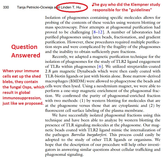
https://www.ncbi.nlm.nih.gov/pubmed/29032556
Tufts. Now saying the fungal Osps are in the blebs, and of course, Osps cause immunosuppression – triacyl lipoproteins cause immunosuppression and never could have been vaccines, and Dearborn is certainly a lie.
============
Questions?
Look for me on Facebook.
Kathleen Dickson
I use the Archangel Michael as an icon.
Stabbing the devil in the head.
Yeah.
Fools.

Reblogged this on and commented:
MUST READ: “Inhibition of Apoptosis” by Kathleen Dickson
LikeLike
Everything I needed to know written in CLAY! I love this article. I’ve been looking for how the macrophages get disabled for some time so one of my listmembers sent me this article were you did all of the work for us two years earlier. I am extremely full of grate.
LikeLike